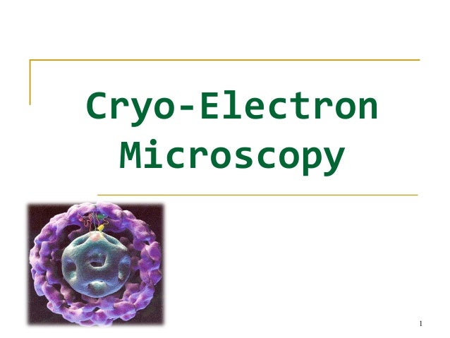
Single Particle Cryo Em

Single Particle Cryo-Electron Microscopy (Cryo-EM) is a powerful technique that has revolutionized the field of structural biology, offering unprecedented insights into the molecular world. This cutting-edge method allows researchers to visualize and study biological molecules, such as proteins, nucleic acids, and their complexes, at near-atomic resolution. By freezing biological samples in a thin layer of ice and capturing high-resolution images, Cryo-EM has become an indispensable tool for understanding the intricate structures and functions of life's building blocks.
The Power of Single Particle Cryo-EM

Single Particle Cryo-EM has emerged as a game-changer in structural biology, providing researchers with an array of advantages over traditional methods. One of its key strengths lies in its ability to analyze heterogeneous samples, a common challenge in studying biological molecules. Unlike X-ray crystallography, which requires a homogenous crystal, Cryo-EM can handle samples with varying conformations, sizes, and orientations. This flexibility makes it an ideal choice for studying dynamic biological processes and complex macromolecular assemblies.
Moreover, Cryo-EM offers an unparalleled resolution, allowing researchers to visualize the atomic details of proteins and other macromolecules. With advancements in technology and image processing algorithms, the technique has pushed the boundaries of resolution, providing structural information at near-atomic levels. This level of detail is crucial for understanding the intricate interactions and mechanisms of biological molecules, leading to breakthroughs in drug discovery, enzyme engineering, and the development of targeted therapies.
The Journey of a Sample in Cryo-EM

The process of Single Particle Cryo-EM involves several intricate steps, each designed to preserve the sample’s integrity and capture high-quality images. It begins with sample preparation, where the biological molecules of interest are carefully purified and concentrated. This step is crucial as it ensures that the sample is free from contaminants and has a high concentration of the target molecule.
Once the sample is prepared, it is applied to a thin layer of carbon or a holey grid, which acts as a support for the sample. The grid is then rapidly frozen in liquid ethane, creating a thin layer of vitreous ice that encapsulates the molecules. This vitrification process is critical as it preserves the sample's structure, preventing ice crystals from forming and causing damage.
With the sample frozen and immobilized, the next step is data acquisition. The grid is loaded into a specialized electron microscope, equipped with a high-sensitivity camera. The microscope beams a focused electron beam onto the sample, capturing thousands of images at various angles and orientations. These images, known as micrographs, contain the structural information of the molecules, which is then extracted and processed to create a 3D reconstruction.
Image Processing and 3D Reconstruction
The image processing stage is a complex and critical step in Cryo-EM. It involves aligning and averaging thousands of individual particle images to enhance the signal and reduce noise. Advanced algorithms and computational power are employed to identify and extract the structural information from the micrographs. This process is often iterative, with multiple rounds of refinement and validation to ensure the accuracy and reliability of the final 3D model.
Once the image processing is complete, the data is ready for 3D reconstruction. Using specialized software, the aligned and averaged particle images are combined to create a 3D density map. This map represents the structure of the molecule, providing a detailed view of its shape, size, and atomic arrangement. The resolution of the final 3D model depends on various factors, including the quality of the sample, the number of particles imaged, and the sophistication of the image processing techniques employed.
| Cryo-EM Resolution | Description |
|---|---|
| Low Resolution (30 Å or higher) | Provides overall shape and basic features. |
| Medium Resolution (10-30 Å) | Allows visualization of secondary structure elements and larger functional sites. |
| High Resolution (3-10 Å) | Reveals atomic details, including side-chain positions and ligand binding sites. |
| Near-Atomic Resolution (3 Å or lower) | Provides near-atomic level detail, allowing accurate modeling of protein structures. |

Applications and Impact
Single Particle Cryo-EM has found widespread applications across various fields of biology and medicine. Its ability to provide high-resolution structural information has accelerated drug discovery efforts, enabling researchers to design targeted therapies with greater precision. By visualizing the binding sites and interactions of proteins with potential drug molecules, Cryo-EM guides the development of novel pharmaceuticals.
In addition, Cryo-EM has revolutionized the study of membrane proteins, which are notoriously difficult to analyze using traditional methods. These proteins, embedded in cellular membranes, play crucial roles in cell signaling, transport, and energy production. With Cryo-EM, researchers can now obtain detailed structures of membrane proteins, opening up new avenues for understanding their functions and developing targeted therapies for membrane-associated diseases.
Furthermore, the technique has expanded our understanding of complex macromolecular assemblies, such as viral capsids and ribosomes. By visualizing these intricate structures at near-atomic resolution, Cryo-EM provides insights into their assembly mechanisms, functions, and potential vulnerabilities. This knowledge is instrumental in developing strategies to combat viral infections and understand fundamental cellular processes.
The Future of Single Particle Cryo-EM
As technology advances, Single Particle Cryo-EM is poised to continue its transformative journey. The development of more powerful electron microscopes, advanced detectors, and sophisticated image processing algorithms will further enhance the technique’s capabilities. Researchers anticipate achieving even higher resolutions, allowing for a deeper understanding of biological structures and their dynamic behavior.
Moreover, the integration of artificial intelligence and machine learning into Cryo-EM workflows holds immense potential. These technologies can automate and optimize various steps of the process, from image processing to 3D reconstruction, making the technique more efficient and accessible. With continued advancements, Single Particle Cryo-EM will likely become an even more versatile and widely used tool in the quest to unravel the secrets of life at the molecular level.
What is the advantage of Single Particle Cryo-EM over other structural biology techniques like X-ray crystallography?
+Single Particle Cryo-EM offers several advantages over X-ray crystallography. It can handle heterogeneous samples, making it ideal for studying dynamic biological processes and complex assemblies. Additionally, Cryo-EM provides near-atomic resolution without the need for crystal formation, which can be challenging for some molecules.
How does the resolution of Cryo-EM compare to other structural techniques?
+Cryo-EM has pushed the boundaries of resolution, achieving near-atomic levels. While X-ray crystallography can also reach high resolutions, Cryo-EM’s ability to handle heterogeneous samples and its recent advancements in technology have made it a powerful tool for obtaining detailed structural information.
What are the challenges and limitations of Single Particle Cryo-EM?
+One of the main challenges is the need for high-quality samples. Preparing samples that are free from contaminants and have a high concentration of the target molecule can be complex. Additionally, the image processing and 3D reconstruction steps require significant computational resources and expertise.


